
Figure 2. Cytotoxicity of cetuximab-IONPs in human GBM neurospheres and U87MGwtEGFR GBM cell line and quantification by an MTT assay.
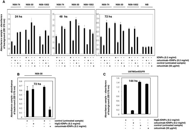
(A) Neurospheres N08-74, N08-30, and N08-1002 (3×10⁴ cells per well) and normal brain cells (NB, 5×10³) were treated with free IONPs (0.2 mg/ml), cetuximab-IONPs (0.2 mg/ml), control vehicle, or cetuximab alone (50 μg/ml) and MTT assay was performed after 24, 48, and 72 hs (GBM neurospheres) or 72 hs (normal brain cells). A significant decrease in cell survival was observed in GBM neurospheres treated with cetuximab-IONPs for 72 hs (P<0.001). No cytotoxicity was observed in normal brain cells after 72 hs. (B) Neurospheres N08-30 were treated with 0.2 mg/ml cetuximab-IONPs or IgG-IONPs for 72 hs when an MTT assay was performed. Only cetuximab-IONPs displayed increased cytotoxicity (P<0.001). (C) U87MGwtEGFR cells (5×10³) were treated with hIgG-IONPs (0.2 mg/ml), cetuximab-IONPs (0.2 mg/ml), control vehicle, or cetuximab alone (50 μg/ml) for 144 hs. A significant decrease in cell survival was found in U87MGwtEGFR GBM cell treated with the cetuximab-IONPs (P<0.001). In all experiments, neurospheres and other cells were used in early passage.