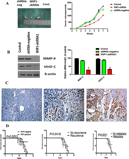
Figure 5. MMP-9 and VEGF-C were downstream targets of WIP1 in vivo and WIP1 expression was associated with the poorer prognosis of ACC patients.

(A), Stable WIP1-shRNA1 ACC-M cells were subcutaneously injected into nude mice. Individual tumor volume was measured at the 7th week after injection and growth curve of xenograft tumors was shown. (B), Western blotting and RT-PCR analysis of the protein and mRNA levels of MMP-9 and VEGF-C in shRNA-neg, WIP1-shRNA1 and control group. Error bars represent the mean ± SD of triplicate experiments (*p < 0.05). (C), WIP1 expression was associated with invasive subtypes of human ACC. Representative images of the immunohistochemical staining of WIP1 in ACC samples. C left, WIP1 in normal human salivary tissue. C middle, WIP1 in weak tumor staining. C right, WIP1 in strong tumor staining. Original magnification, × 100; inset, × 200; bar, 100 mm. (D), Kaplan-Meier survival analysis in patients with ACC. Overexpression of WIP1 in ACC was associated with a shorter overall survival in the respective group.
