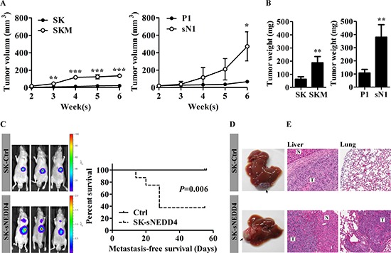
Figure 4. sNEDD4 promotes tumorigenesis and metastasis in vivo.

(A) SK, SKM, P1 and sN1 cells were subcutaneously injected into the flanks of nude mice, respectively, and tumor volumes measured once a week (n = 4 for each group). Tumor growth curves are shown. (B) Six weeks later, tumors were excised and weighed. C–E, Tumors dissected from mice subjected to subcutaneous injection with control and SK-sNEDD4 cells were introduced orthotopically into nude mice and monitored weekly with IVIS. (C) left, Representative IVIS images of mice 5 weeks after tumor implantation (9 mice/group). The metastasis-free survival curves are shown (C, right). Mice were sacrificed 8 weeks after implantation. Livers (the implantation site is highlighted with arrows in panel D) and lungs were excised, fixed and embedded in paraffin. Hematoxylin & Eosin (H&E) staining was performed on liver (E, left) and lung (E, right) tissue sections. N, normal; T, tumor; *P < 0.05; **P < 0.01; ***P < 0.001.
