
Figure 5. Human and mouse Shp2 have differential effects on EGF-stimulated ERK activation as a result of different SUMOylation levels.
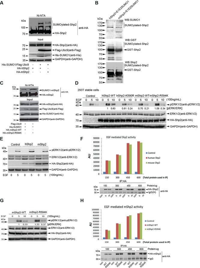
(A) In vivo Ni2+-NTA resin pull-down SUMOylation assays for human and mouse Shp2 in 293T cells. (B) In vitro GST agarose pull-down SUMOylation assays for human and mouse Shp2 with pE1E2SUMO1 into E.coli BL21 (DE3). (C) SUMOylation assays for mouse Shp2-WT and -R594K mutant in 293T cells by using the method of Ni2+-NTA resin pull-down. (D) Immunoblotting analysis of ERK1/2 phosphorylation of stable 293T cell lines expressing hShp2WT, hShp2K590R, mShp2WT and mShp2R594K. (E–F) Serum-starved 293T cells transiently expressing hShp2 or mShp2 were stimulated with 100 ng/mL of EGF for 5 min and the ERK activities were determined by Western blotting (E); the same lysates were used for immunoprecipitation with anti-HA, then performed the Shp2 phosphatase activity assays (F). (G–H) Serum-starved 293T cells transiently expressing mShp2WT or mShp2R594K were stimulated with 100 ng/mL of EGF for 5 or 10 min and the ERK activities were determined by Western blotting (G); the same lysates (5 min) were used for immunoprecipitation with anti-HA, then performed the Shp2 phosphatase activity assays (H).