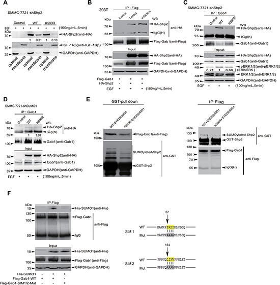
Figure 6. SUMOyaltion of Shp2 promotes ERK activation by controlling its association with Gab1.

(A) Cytosolic fractions and membranous fractions extracted from EGF-stimulated SMMC-7721-shShp2 cells stably re-expressing hShp2WT or hShp2K590R were analyzed by Western blotting with antibodies against HA, IGF-1Rβ (as a membrane protein marker) and GAPDH (as a cytosolic marker). (B) 293T or 293T-shSENP1 cells were co-transfected with HA-Shp2 and Flag-Gab1 plasmids. 24 h after transfection, cells were subjected to serum deprivation for 16 h, followed by the treatment with 100 ng/mL of EGF for 5 min. Cell lysates were immunoprecipitated and subsequently immunoblotted with indicated antibodies. (C) Lysates from EGF-treated SMMC-7721-shShp2 cells stably re-expressing hShp2WT or hShp2K590R were immunoprecipitated with anti-Gab1 antibody and then immunoblotted with anti-HA antibody. Lysates were also used as Input for immunoblotting with antibodies against pERK1/2 and other indicated. (D) Lysates from EGF-treated SMMC-7721-shSUMO1 cells stably re-expressing hShp2WT or hShp2K590R were immunoprecipitated with anti-Gab1 antibody and then immunoblotted with anti-HA antibody. Lysates were also used as Input for immunoblotting with antibodies indicated. (E) Flag-Gab1 was transiently expressed in 293T cells and GST-Shp2 with pE1E2SUMO1 were transformed into E.coli BL21 (DE3). Two reciprocal pull-down assays of GST-proteins (left panels) and anti-Flag/IP (right panels) were performed, and then immunoblotted. (F) Lysates from 293T cells co-transfected with His-SUMO1 and Flag-Gab1WT or Flag-Gab1SIM1/2mut palsmids were immunoprecipitated with anti-Flag antibody, and then immunoblotted with anti-His antibody. Lysates were also used as Input for immunoblotting with indicated antibodies (left panels). The consensus amino acid sequences (yellow labeled) of predicted SIM1 and SIM2 of Gab1 were mutated to alanine (right panels).
