
Figure 6. Bafetinib treatment impairs the formation of breast cancer liver metastases.
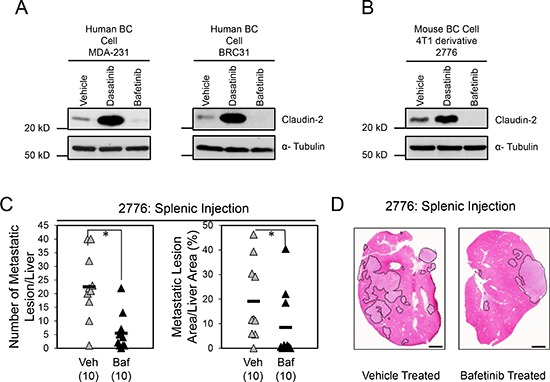
Immunoblot analysis of Claudin-2 expression in both human breast cancer cells (MDA-MB-231 and BRC31) (A) or 2776 mouse breast cancer cells (B) following treatment with Bafetinib inhibitor. As a loading control, total cell lysates were blotted for α-Tubulin (A, B). (C) Bafetinib treatment decreases the formation of liver metastases derived from 2776 cells following splenic injection (1 × 10⁵ cells). A statistically significant decrease in both the number of hepatic metastatic lesions or the liver metastatic burden is observed when the control cohort (Par Vehicle) is compared to the Bafetinib-treated cohort (Par Baf) (*, P < 0.05). The number of mice analyzed in each cohort is indicated in parentheses. (D) Representative images of H&E stained liver sections exhibiting the liver metastatic burden in each cohort. Scale bar represents 2mm. Veh, Vehicle; Baf, Lyn inhibitor (Bafetinib).