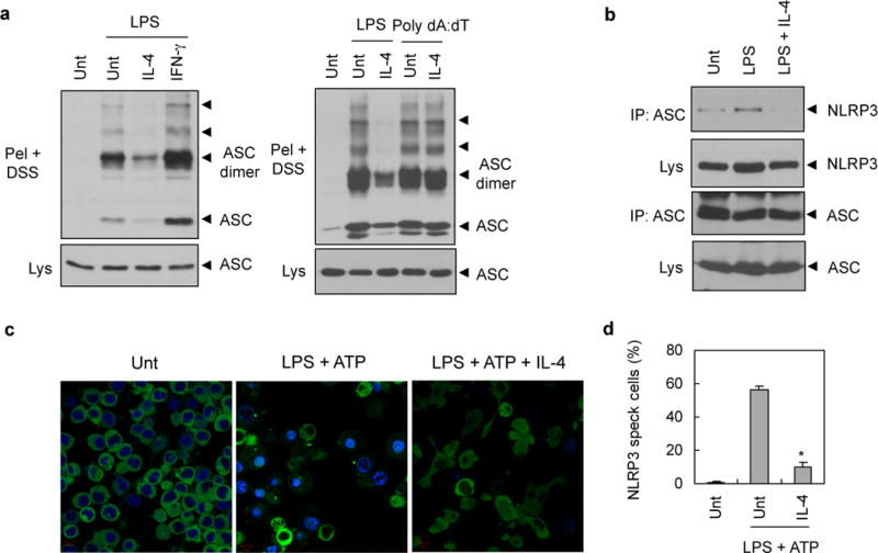
Figure 3. Inhibition of NLRP3 inflammasome assembly by IL-4.

(a) PMA-differentiated THP-1 cells were untreated (Unt) or treated with LPS (0.5 μg ml−1, 6h) or transfected with poly dA:dT (2 μg, 6h) in the presence or absence of IL-4 or IFN-γ pretreatment (20 ng ml−1, 3h). Chemical crosslinking was then performed as described in Materials and methods. DSS-crosslinked NP-40 insoluble pellets (Pel + DSS) or soluble lysates (Lys) were immunoblotted with anti-ASC antibody. (b) PMA-differentiated THP-1 cells were untreated (Unt) or treated with LPS (0.5 μg ml−1, 3h) in the presence or absence of IL-4 pretreatment (20 ng ml−1, 3h). Soluble lysates were immunoprecipitated with anti-ASC antibody, and the immunoprecipitates were then immunoblotted with anti-NLRP3 antibody. (c) NLRP3-GFP-expressing BMMs were untreated (Unt) or treated with LPS (0.25 μg ml−1, 3h) in the presence or absence of IL-4 pretreatment for 3h (20 ng ml−1), followed with ATP (2.5 mM, 30 min). Cells were then observed by confocal microscope. Scale bar, 10 μM. (d) Relative percentages of NLRP3-speck containing cells as treated in Fig. 3c. Asterisk indicates significant difference from LPS plus ATP-treated sample. (n = 3, *P<0.005)
