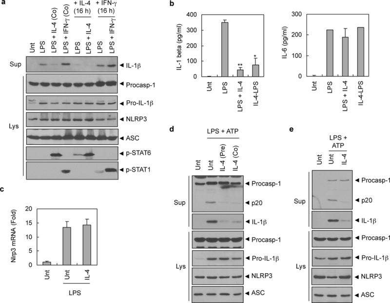
Figure 4. IL-4 inhibits NLRP3 inflammasome in a M2 polarization- and NLRP3 transcription-independent manner.

(a) PMA-differentiated THP-1 cells were untreated (Unt) or pretreated with IL-4 (lane 5, 6) or IFN-γ (lane 7, 8) (20 ng ml−1) for 16h, and then stimulated with LPS (0.5 μg ml−1) together with IL-4 (lane 3, 6) or IFN-γ (lane 4, 8) for 6h. Culture supernatants or lysates were immunoblotted with the indicated antibodies. (b) PMA-differentiated THP-1 cells were untreated (Unt) or treated with LPS (0.5 μg ml−1) alone (LPS) or together with IL-4 (20 ng ml−1, LPS+IL-4) for 6h, or pretreated with IL-4 (20 ng ml−1) for 16h before LPS stimulation (IL-4-LPS). Culture supernatants were assayed for extracellular IL-1β or IL-6 by ELISA. The asterisks indicate significant differences from LPS-treated cells. (n = 2, *P<0.05; **P<0.01) (c) Murine BMMs were untreated (Unt) or treated with LPS (0.25 μg ml−1, 3h) in the presence or absence of IL-4 pretreatment (20 ng ml−1, 3h). The mRNA level of Nlrp3 was determined by quantitative real-time PCR. Three independently treated wells were analyzed. (d) Murine BMMs were untreated (Unt), or treated with LPS (0.25 μg ml−1, 3h) in the presence or absence of IL-4 pretreatment (pre) for 3h followed with ATP (3 mM, 45 min), or treated with LPS (0.25 μg ml−1, 3h) followed with co-treatment of ATP and IL-4 (Co; 20 ng ml−1) for 45 min. (e) NLRP3-reconstituted Nlrp3-knockout macrophages (N1–8) were untreated (Unt) or treated with LPS (0.25 μg ml−1, 3h) in the presence or absence of IL-4 pretreatment (20 ng ml−1, 3h) followed with ATP (3 mM, 45 min). (d–e) Culture supernatants (Sup) or lysates (Lys) were immunoblotted with the indicated antibodies.
