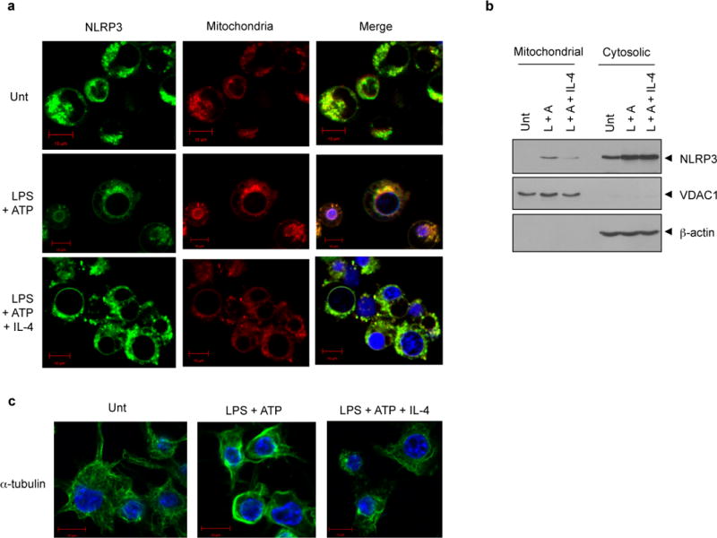
Figure 6. IL-4 modulates the subcellular localization of NLRP3 and microtubule polymerization.

(a) NLRP3-GFP-expressing BMMs were untreated (Unt) or primed with LPS (0.25 μg ml−1, 3h) in the presence or absence of IL-4 pretreatment (20 ng ml−1, 3h), followed with ATP (2.5 mM, 30 min). Cells were stained with MitoTracker and observed by confocal microscope. Scale bar, 10 μM. (b) Murine BMMs were treated as in (a) and the cell lysates were fractionated into cytosolic and mitochondrial fraction, and then immunoblotted with the indicated antibodies. (L, LPS; A, ATP) (c) Murine BMMs were treated as in (a) and stained with the anti-α-tubulin antibody. Then, the immunofluorescence assay was performed as described in Materials and methods. Blue signals represent nuclear fluorescence. Scale bar, 10 μM.
