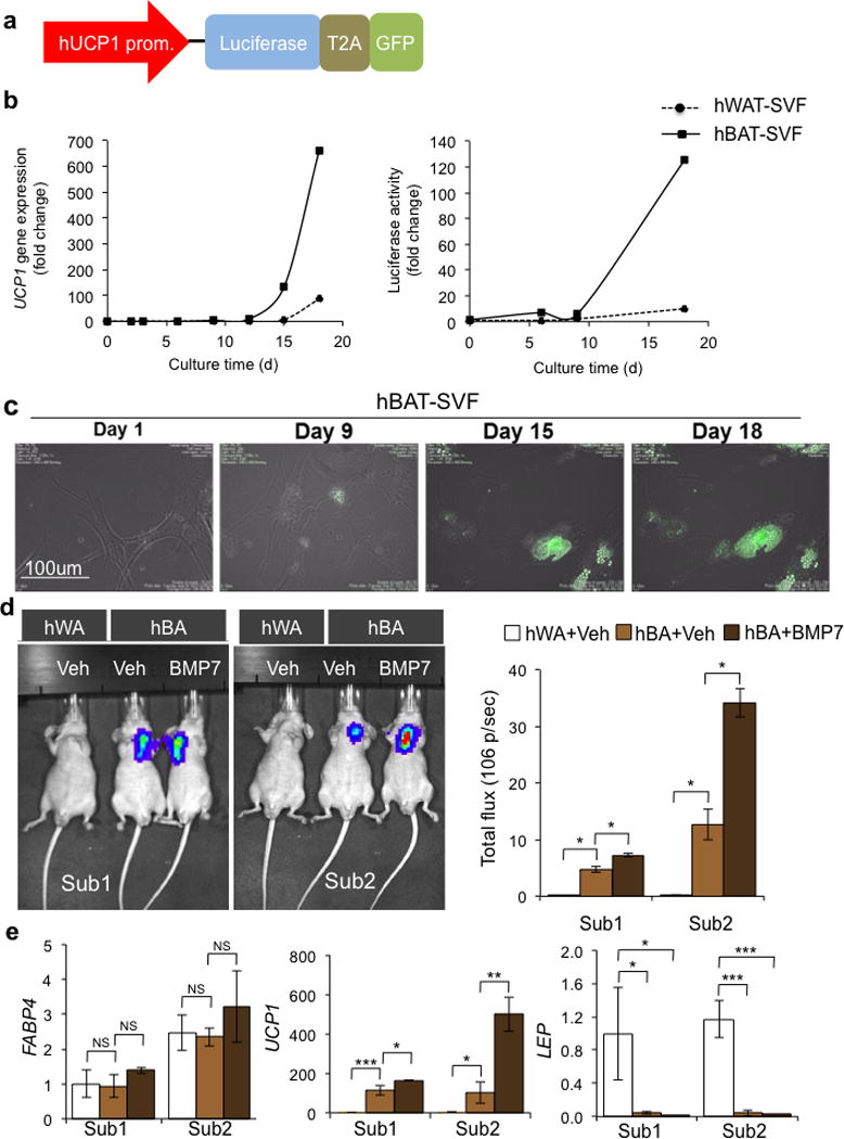
Figure 2.

Utilization of a UCP1 reporter system for in vitro and in vivo monitoring of UCP1 expression. (a) Schematic structure of the hUCP1 promoter reporter system. 4148 bp of human UCP1 promoter drives the expression of bicistronic luciferase and GFP. T2A is the internal ribosomal entry site. (b) In hBAT-SVF and hWAT-SVF stably expressed the reporter construct, luciferase activity (Right) was strongly correlated with endogenous UCP1 gene expression (Left) during the course of differentiation (see Fig. 1a and Method). Data are presented as fold changes compared to hWAT-SVF on day 0 (mean ± s.e.m., n=3). A representative experiment from a total of two independent studies is shown. (c) Monitoring UCP1 expression by GFP in vitro using a time lapse imaging system during differentiation of hBAT-SVF from Sub1. (d) Representative IVIS images of nude mice after 22 days of transplantation of hWAT-SVF and hBAT-SVF are shown on the left panel. Quantifications of luciferase activity by total flux are shown on the right panel (mean ± s.e.m.). The experiments have been repeated twice (n=2 for hWAT-SVF group; n=3 for hBAT-SVF group). (e) Q-RT-PCR analysis for expression of FABP4, UCP1 and LEP in fat pads developed from the transplanted cells. Data are presented as fold changes compared to fat pads developed from hWAT-SVF with vehicle treatment (mean ± s.e.m.). Two-tailed Student’s t-test was used to determine P values (* P < 0.05, ** P < 0.01, *** P < 0.001).
