
Figure 3.
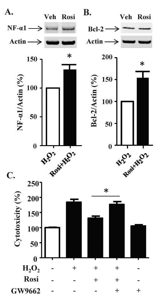
PPARγ mediates the protective effect of Rosi in Neuro2a cells. (A, B) Neuro2a cells were incubated with vehicle or Rosi for 24 h, then the cells were treated with 100 μM H2O2 for 24 h. Western Blots showed (A) NF-α1 and (B) BCL-2 protein levels increased with Rosi treatment (see Fig. 2) and remained elevated after 100 μM H2O2 treatment in Neuro2a cells. n = 3/group; values are mean ±SEM, p<0.05, t-test. (C) Neuro2a cells were treated with 1 μM Rosi and/or 1 μM GW9662 for 24 h and then the cells were treated with 100 μM H2O2 for 24 h. The LDH release assay showed that GW9662 inhibited the protective effect of Rosi against oxidative stress in Neuro2a cells. n=5/ group; values are mean ±SEM, one way ANOVA for treatment effect: F(4, 20)=93.69, p<0.001, followed by Tukey test. *p <0.05, GW9662 + Rosi + H2O2 group compared to Rosi + H2O2 group.