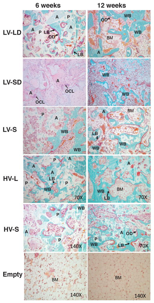
Figure 5. High-magnification images of histological sections of defects treated with injectable allograft/polymer composites.
All of the images show sections stained with Goldner’s trichrome except for LV-SD at 6 weeks, which is stained with H&E. Original magnification 70X. With Goldner’s trichrome stain: white- residual polymer (P); light blue particles with angled shapes- residual allograft particles (A); blue with cells inside- new mineralized bone with osteocytes with a woven structure (WB); red- an organized lamellar bone structure (LB); red bone lining cells- osteoid (OD); and bone marrow (BM). Arrows in the LV-SD 6 weeks section point to multi-nucleated osteoclast-like cells (OCLs).

