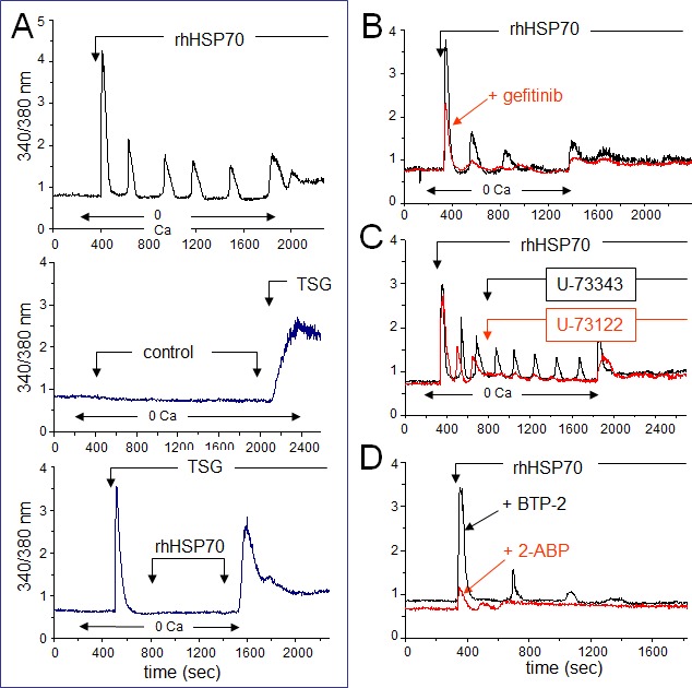
Fig 4. Extracellular rhHSP70 induces intracellular Ca2+ mobilization.

A. rhHSP70 induced Ca2+ release from internal stores in HMEC. Data are expressed as the 340/380 nm excitation ratio in one cell to observe oscillations in [Ca2+]i because the oscillatory process is not synchronized in cells of the same monolayer. External additions of drugs are indicated by arrows. Changes in external calcium bath conditions are indicated on the bottom of traces. In most cases, drugs were initially applied in the absence (0 Ca) then in Ca2+ (1.8 mM) containing solution to reveal Ca2+ release from internal stores then external Ca2+ entry, respectively (representative from 50 cells; n=10). No calcium increase was induced by the cell superfusion of the control bath solution (middle trace) while thapsigargin (TSG 4μM) always produced a drastic increase in [Ca2+]i (lower trace; Representative from 50 cells; n=4). B. Contribution of EGFR to rhHSP70-induced Ca2+ signaling. Superimposed traces from cells preincubated with the EGFR (ErbB1) inhibitor, gefitinib (10 μM for 30 min; in red), before the addition of rhHSP70 (Representative from 50 cells; n=5). C. Effects of phospholipase C (PLC) inhibitor U-73122 (5 μM) and its inactive analog U-73343 (5 μM) on the rhHSP70-induced Ca2+ oscillations. Drugs were applied without (0 Ca) then with extracellular Ca2+ (1.8 mM) (representative from 50 cells; n=5). D. Ca2+ oscillations required both Ca2+ release from internal stores and store operated Ca2+ entry (SOCE). Cells were pretreated with the selective SOCE inhibitor, BTP-2 (20 μM; 20 min), before challenged with rhHSP70 (Representative from 30 cells; n=4).
