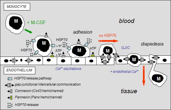
Fig 8. Hypothetical model of the inhibitory effects of extracellular HSP70 on the diapedesis of monocytes.

The diagram shows the distribution of gap-junctions (double barrel) and hemichannels (single barrel) that can operate between monocytes (M) and endothelial cells of the microvascular wall. M-CSF-stimulated monocytes adhere to the endothelial cell monolayer and release HSP70 that disrupts GJIC between EC contributing to the subsequent release of ATP through Panx-1 and Cx43 hemichannels from endothelial cells. Extracellular HSP70 induce endothelial Ca2+ oscillations without affecting the intercellular tight junction protein, localized between apposed cells. When HSP70 is blocked (siRNA or VER155008), M-CSF-stimulated monocytes communicate with endothelial cells by gap junctions (GJIC), allowing their migration across the endothelial cell monolayer, in an endothelial Ca2+-dependent mechanism (slowed down by BAPTA, a Ca2+ chelator, Suppl. Fig. S5).
