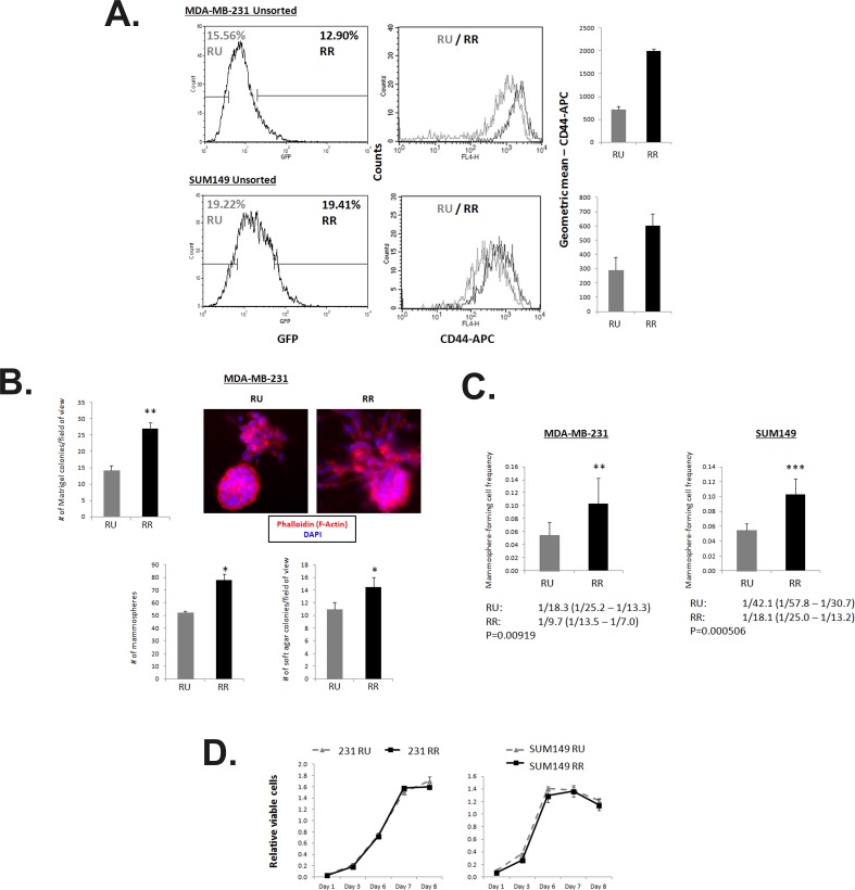
Figure 3. RR cells exhibit higher CD44 expression, enhanced capacities for colony formation in vitro and higher frequency of mammosphere-forming cells.
A. Flow cytometry analyses of MDA-MB-231 and SUM149 Unsorted SRR2 cells stained with CD44-APC. Cells were gated on the highest and lowest 10 to 20% GFP expression and analyzed for CD44-APC levels. B. Results for Matrigel colony formation assay, conventional mammosphere assay, and soft agar assay of untreated MDA-MB-231 RU and RR cells are shown. 2500 cells/well are seeded into a 96-well Matrigel colony formation assay and colonies are counted from photographs taken on Day 7. Photographs of Matrigel multi-cell colonies were stained with phalloidin and imaged by high content screening imaging microscopy. 10,000 cells/well are seeded into a 6-well mammosphere assay and counted on Day 7. 10,000 cells/well are seeded into a 24-well soft agar assay and counted on Day 28. C. Extreme limiting dilution analyses statistics and graphical depiction of results are shown of a limiting dilution mammosphere assay in a 96-well plate format. Cells were seeded in 10 seeding densities ranging from 1 to 1000 cells/well in 6 replicates each. D. MTS 2-dimensional proliferation assay quantification of untreated ER− RU and RR cells seeded at 2000 cells/well. 20 μL of MTS reagent is added with fresh media 2 hours prior to taking absorbance reading.

