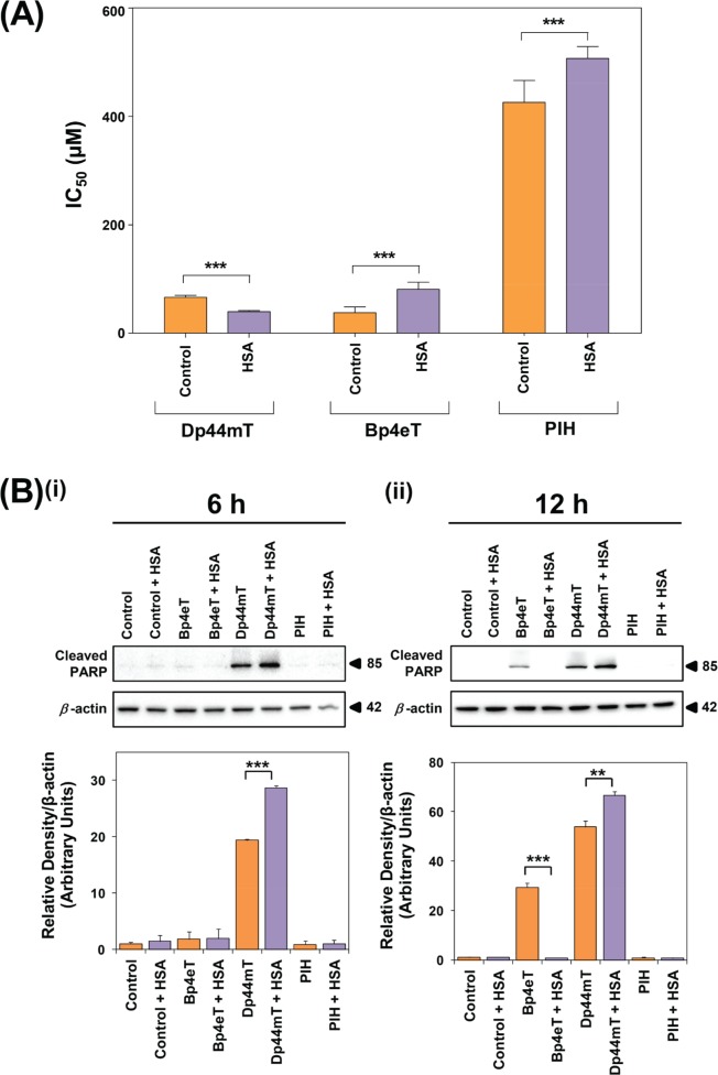
Figure 6. The effect of HSA on the anti-proliferative and apoptotic activity of Dp44mT, Bp4eT and PIH.
(A) The anti-proliferative activity of Dp44mT, Bp4eT and PIH in the presence of HSA for 24 h/37°C. Cells were incubated with Dp44mT (30-120 μM), Bp4eT (30-120 μM), PIH (150-600 μM) or vehicle alone (control) in the presence or absence of HSA (40 mg/mL) for 24 h/37°C. Trypan blue was used to obtain direct cell counts and to determine IC50 values. Results are expressed as mean ± S.E.M. (3 experiments). ***, p<0.001 relative to the corresponding control. (B) Levels of cleaved PARP following treatment of SK-N-MC cells with Bp4eT, Dp44mT or PIH (50 μM) in the presence and absence of HSA (40 mg/mL) after (i) 6 h or (ii) 12 h/37°C. Western blots are typical of 3 independent experiments. Results are expressed as mean ± S.E.M. (3 experiments). **, p<0.01; ***, p<0.001.

