
Figure 2. iVR1 inhibited growth and neo-angiogenesis of syngenic and xenograft colorectal tumors.
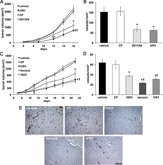
CT26 mouse colon carcinoma cells (A) or HCT-116 human colorectal cancer cells (C) were injected subcutaneously in Balb/c mice or CD1 nude athymic mice, respectively. iVR1 and control peptide (CP) were delivered at 50 mg/kg each other day. Anti-PlGF mAbs 5D11D4 (mouse) and 16D3 (human) were delivered at 25 mg/kg twice a week. Bevacizumab (bevaciz) was delivered at 5 mg/kg, twice a week. Vehicle was PEG400/water 1:1. TV was measured three times a week and data are represented as the mean ± SEM (N = 7). A, *p < 0.001 and §p < 0.0001 versus vehicle and CP; ^p < 0.02 and ¶p < 0.05 vs 5D11D4; #p < 0.002 versus vehicle and CP. C, §p < 0.01 and *p = 0.0001 versus vehicle and CP, ¶p < 0.05 versus 16D3; #p = 0.0027 versus vehicle and CP. Vessel density of syngenic (B) and xenograft (D) tumors, were calculated analyzing five optical fields for each tumor, counting CD31-positive vessels (brown). Data are represented as the mean ± SEM. B, *p < 0.007 versus vehicle and CP. D, §p < 0.005, #p < 0.02, and *p < 0.0005, versus vehicle and CP. ¶p < 0.05 bevaciz versus iVR1 and CP and iVR1 versus 16D3. (E) Representative pictures of CD31 staining (brown) of HCT-116 tumors. Scale bar, 100 μm.