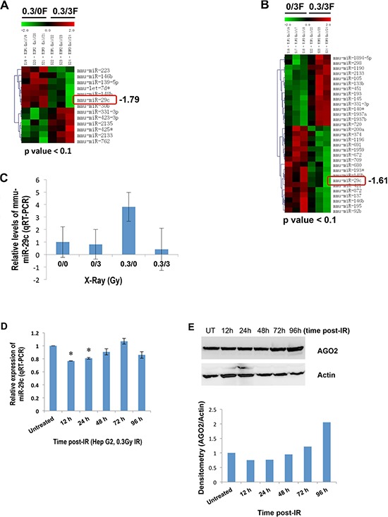
Figure 1. A differential expression of miR-29c in liver tissues of female mice exposed to IR and human HepG2 cells.

(A and B) Total RNA isolated from IR-exposed female mouse liver tissues 96 hours post-irradiation was subjected to the microRNA microarray. (C) Total RNA was isolated from liver tissues of female mice exposed to IR 96 hours post irradiation, and the levels of miR-29c were examined by real-time RT-PCR. (D) Total RNA isolated from human hepatocellular carcinoma HepG2 cells at the indicated time points after exposure to low-dose X-ray (0.3 Gy) was subjected to real-time RT-PCR using miR-29c primer. (E) Whole cellular lysate prepared from HepG2 cells at the indicated time points after exposure to low-dose X-ray (0.3 Gy) was subjected to Western blotting with a specific antibody to AGO2. The asterisk indicates p < 0.05.
