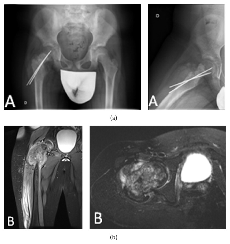
Figure 1.

(a) Plain radiographs of the pelvis, hip, and proximal femur showing mixed osteolytic and sclerotic areas in the right proximal femur and an unhealed femoral neck fracture fixed with two K wires. (b) MRI showing a proximal femoral lesion extending distally 13 cm with a surrounding soft-tissue mass.
