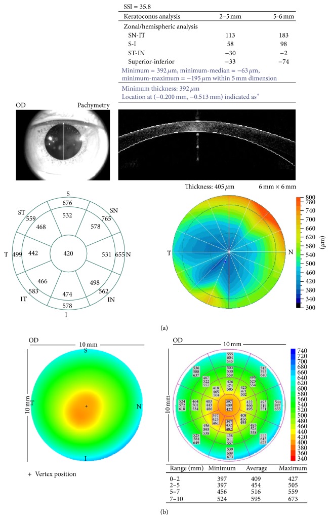
Figure 1.
Corneal pachymetry of a patient with keratoconus generated by the (a) RTVue-OCT and (b) Visante-OCT. (a) Minimum of corneal thickness can be acquired from the table of “keratoconus analysis” on the upper right and the thinnest point is also marked on the pachymetric map on the lower right. A real-time monitoring on the upper left is used for pupil centering. (b) Minimum of corneal thickness in different regions is demonstrated on the table on the lower right and the thinnest point is also marked on the pachymetric map on the left.

