Figure 6.
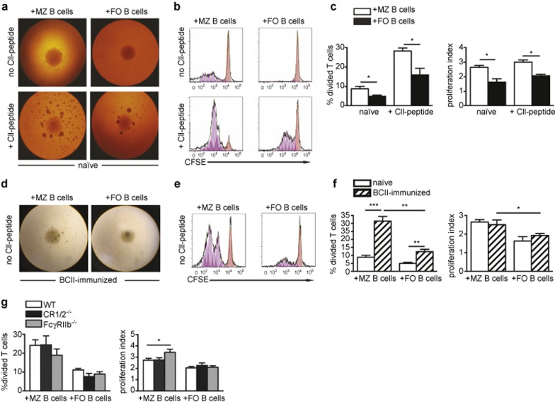
MZ B cells are superior to FO B cells in presenting autoantigen to T cells and show enhanced capacity when deficient in FcγRIIb. CFSE-labeled Vβ8.3+ CII-specific T cells were cultured with splenic MZ or FO B cells from naïve or BCII-immunized mice, with or without addition of CII(245-270)-peptide. (a) Cultures of Vβ8.3+ CII-T cells and B-cell subsets from naïve mice at 3 days with or without CII-peptide (×4 magnification). (b) Cultures in a shown as histograms of CFSE dilution. The orange peaks show undivided cells, the pink peaks show generations of cells in division. (c) Percentage of CII-specific T cells that went into division (left) and the proliferation index of the dividing T cells (right) after culture as described in a (n = 4). Data were presented as mean + SEM and represent three independent experiments. (d) Cultures of Vβ8.3+ CII-specific T cells and B-cell subsets from BCII-immunized mice (×4 magnification). (e) Cultures in d shown as histograms of CFSE dilution. The orange peaks show undivided cells, the pink peaks show generations of cells in division. (f) Percentage of CII-specific T cells that went into division (left) and the proliferation index of the dividing T cells (right) after culture as described in a and d (n = 4). Data were presented as mean + SEM and represent five independent experiments. (g) Percentage of CII-specific T cells that went into division (left) and the proliferation index of the dividing T cells (right) after culture with MZ or FO B cells from the spleen of BCII-immunized WT-, CR1/2-, or FcγRIIb-deficient mice (n = 5–9). Data were presented as mean + SEM and represent seven independent experiments.

