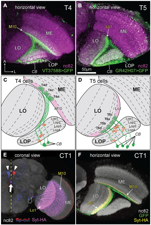
Figure 2.

The morphology of T4, T5, and complex tangential cell (CT1) cells. (A,B) Innervation patterns of T4 and T5 cells. VT37588-Gal4 driven Green Fluorescent Protein GFP(T2), mCD8::GFP (LL6) highlights the dendritic zone in ME stratum M10 for T4 dendrites and GR42H07-Gal4 driven GFP(T2), mCD8::GFP (LL6) highlights the dendritic zone in LO stratum Lo1 for T5 dendrites. Cell bodies (CB) intermingle in the cortex of the LOP; ME: medulla; LO: lobula. Background immunolabel: nc82 (anti-BRP, magenta). (C,D) Innervation patterns of single T4 and T5 cells (after Fischbach and Dittrich, 1989). The presynaptic (output) sites of the cells in the LOP are indicated in orange. Strata housing the dendritic (input) arbors are highlighted in pink [M10 in (C) and Lo1 in (D)]. In each cell type, four subtypes (a, b, c and d) each target one of the LOP’s four strata, Lop1, Lop2, Lop3 and Lop4. Only three T4 subtypes are shown in (C), as originally illustrated by Fischbach and Dittrich (1989). (E) Coronal projection of the CT1 cell. A bilateral pair of CB lies one each on either side of the midline (yellow dashed line), somewhat dorsal to the antennal lobes (white arrowheads). The axons project contralaterally, crossing each other at the midline (arrow) and medial to the optic lobe they bifurcate (black arrowhead) into a ME and a LO fiber. Columnar terminals are visible as a repeated array in strata M10 and Lo1. Blue and red: single cell flip-out clone for each CT1 cell (Nern et al., 2015); magenta: synaptotagmin-hemaglutinin (Syt-HA) indicating presynaptic sites; background immunolabel: nc82 (gray). (F) Innervation of CT1 in the optic lobe, seen in a horizontal plane. A single CT1 cell innervates the M10 and Lo1 strata, both in their entirety, communicating with T4 and T5 cells, respectively. Its axons run over the surface of the neuropils and project into them. A presynaptic marker Syt-HA expresses throughout both strata. Green: CT1-Gal4 driven GFP; yellow: Syt-HA; background immunolabel: nc82 (gray).
