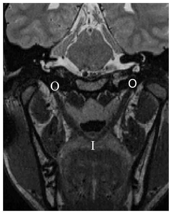
FIGURE 1.

Magnetic resonance imaging in the oblique coronal plane displaying the levator muscle from the origin to the insertion in the velum. “I” represents the insertion of the levator muscle into the velum, and “O” represents the origin at the base of the skull. Note that the muscle forms a sling through the middle of the velum.
