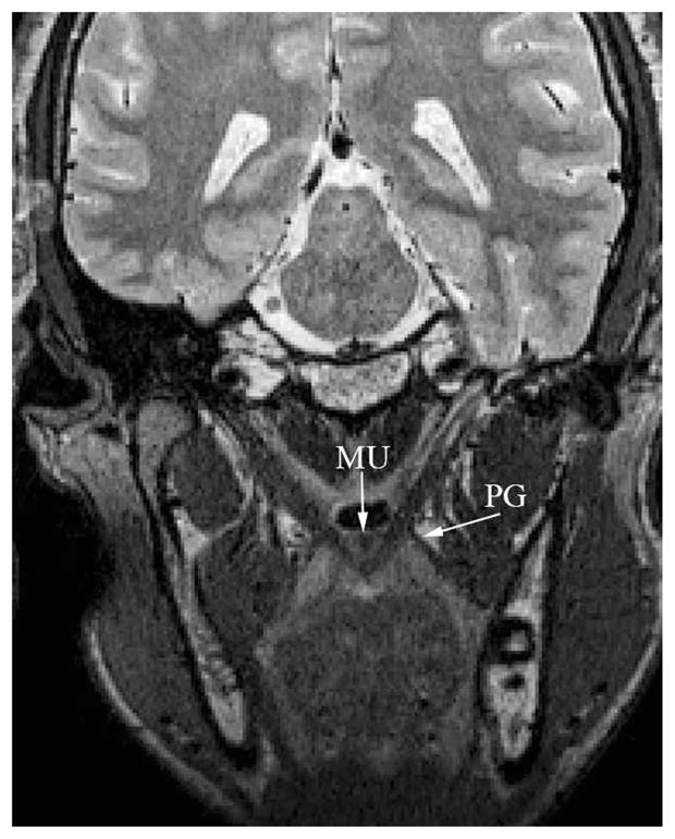
FIGURE 7.

Image of the musculus uvulae (“MU”) on an oblique coronal image plane. Note that the muscle appears near the nasal surface of the velum and is clearly delineated from the levator muscle bundle through the midline of the velum. The palatoglossus (PG) is also evident in the image, and the boundary, as it joins the levator muscle, can be visualized.
