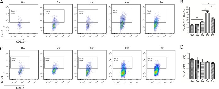
FIG 5.
Increased Tim-3 expression on CD11b+ cells in S. japonicum-infected mice. HMCs (A and B) and splenic immune cells (C and D) from S. japonicum-infected mice were collected at 0, 2, 4, 6, and 8 weeks postinfection, and Tim-3 expression on CD11b+ cells was detected by flow cytometry. (A and C) Representative dot plots of Tim-3 expression on CD11b+ cells. The dot plots are representative of three independent experiments with five to seven mice in each group per experiment. (B and D) Comparison of the proportions of Tim-3+ cells within the CD11b+ cell population among groups of mice at 0, 2, 4, 6, and 8 weeks postinfection (means ± SD; *, P < 0.05; **, P < 0.01; ***, P < 0.001).

