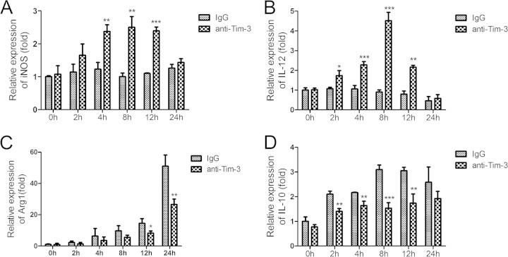
FIG 6.
Tim-3 blockade polarized macrophages from the M2 to M1 phenotype. CD11b+ cells were isolated from HMCs of S. japonicum-infected mice euthanized at 6 weeks postinfection. Anti-Tim-3 antibody was used to block the Tim-3 pathway following SEA treatment, and rat IgG was used as a control. Cells were collected at 0, 2, 4, 6, and 8 h. Macrophage activation-related genes were quantified using real-time RT-PCR. (A) iNOS; (B) IL-12; (C) Arg1; (D) IL-10. Gene expression was normalized against β-tubulin and is presented as the fold change versus the expression of IgG group at week zero. The results are representative of two independent experiments (means ± SD; *, P < 0.05; **, P < 0.01; ***, P < 0.001).

