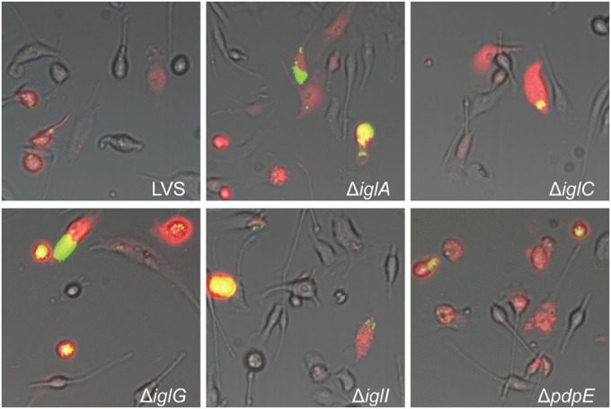
FIG 3.

Microinjection of indicated F. tularensis strains into BMDM. Pictures were taken at 24 h after injection with a live-cell imaging microscope equipped with an EMCCD camera. Colocalization of injected cells containing RD (red) and GFP-expressing bacteria (green) resulted in yellow signals. Representative pictures for each strain from at least three independent experiments are shown.
