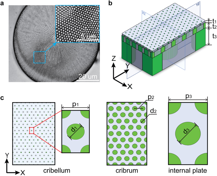
Figure 1.
(a) Optical microscopy images of Coscinodiscus sp. (b) Simplified 3D structure of the unit cell of diatom frustule based on experimental results. Thickness of the three layers: Cribellum t1 = 50 nm, Cribrum t2 = 300 nm, Internal Plate t3 = 1000 nm, (c) Left: top view of cribellum, the lattice constant p1 = 200 nm and the hole size d1 = 50 nm. Middle: top view of cribrum, the lattice constant p2 = 400 nm and the hole size d2 = 250 nm. Right: top view of internal plate, the lattice constant p3 = 2 μm and the hole size d3 = 1.3 μm.

