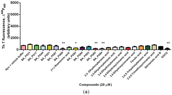
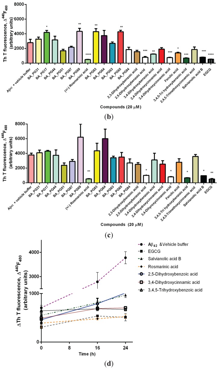
Figure 1.
Amyloid formation assayed by thioflavin T fluorescence. (a) Fibril formation of Aβ42 and the associated increase in in situ ThT fluorescence in presence and absence of compounds were measured at 0 h (baseline) (b) after 16 h (c) and after 24 h incubation; (d) Selected phenolic compounds that resulted in a significant anti-amyloidogenic property compared with the positive control (EGCG) and negative control (Aβ42 & vehicle buffer) determined by ThT measured at 0, 16, and 24 h after incubation with chemically synthesized Aβ42. Significant differences are indicated: * p < 0.05; ** p < 0.01; *** p < 0.005; **** p < 0.001.


