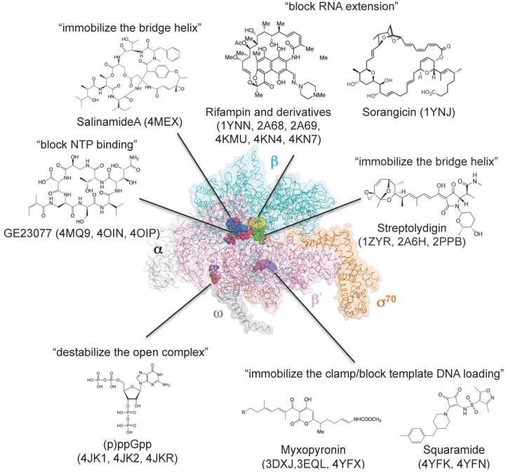
Figure 3.
Three-dimensional representation of targets of small-molecule RNAP inhibitors. E. coli RNAP holoenzyme is depicted as an α-carbon backbone traced with partially transparent molecular surfaces (α subunits: white; β subunit: cyan; β' subunit: pink; ω subunit: gray; σ70: orange). Small-molecule inhibitors bound to RNAP are depicted as CPK models. Chemical structures of small-molecule inhibitors and mechanisms of transcription inhibition are indicated.

