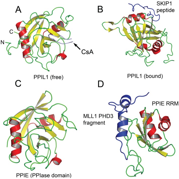
Figure 2.
Structures of PPIL1 and PPIE free and complexed to spliceosomal proteins. In (A), the crystal structure of the free PPIase domain of PPIL1 is shown. The protein has a typical cyPA-like fold; In (B) the solution NMR structure of PPIL1 PPIase domain bound to the SKIP1 peptide is depicted. The SKIP1 peptide forms a hook like structure (in blue) and binds the PPIase domain at an allosteric site far removed from the active site; In (C), the crystal structure of the PPIase domain of PPIE is shown; In (D), the solution NMR structure of the MLL1-PHD3-PPIE-RRM complex is shown. The PHD3 fragment forms a helix that packs against the PPIE RRM.

