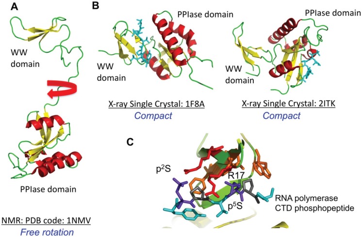
Figure 4.
NMR and X-ray crystal structures of Pin1 free and bound to peptides are shown. In (A), the solution NMR structure (PDB code 1NMV) is depicted showing the Pin1 PPIase domain and the WW domain separated by a linker; In (B) two crystal structures of Pin1 bound to phosphopeptides are shown. In the first structure (PDB code 1F8A), the peptide interacts with the WW domain and in the second complex (PDB code 2ITK), the peptide interacts with the PPIase domain; In (C), the interactions of the WW domain with a doubly phosphorylated Ser-Pro peptide is shown. The phosphoserines are shown in blue and the arginine side chains from the WW domain are shown in red.

