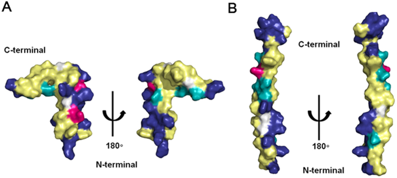
Figure 10. Comparison of the hydrophobic and electrostatic potentials of papiliocin and moricin.

(A) Hydrophobic and electrostatic potentials of papiliocin (pdb: 2LA2) and (B) moricin (pdb: 1 × 22), as determined by NMR spectroscopy. The residues are color-coded as follows: yellow, hydrophobic; cyan, hydrophilic; blue, positive; red, negative; white, neutral.
