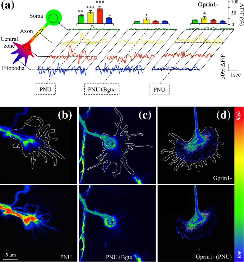
Fig. 6.
Detection an α7 calcium current in the growth cone. Cells were analyzed for intracellular calcium changes using Fluo-8AM. (a) Calcium detection in soma, axon, central zone, and filopodia of hippocampal neurons. Normalized traces were obtained from calcium reading in 10 cells (n=10) where 0.1% DMSO alone was used as a vehicle control. An empty pRNAT H1.1 vector was used as a transfection control for Gprin1-traces. Histograms showing relative changes in calcium peaks at time of drug treatment. (b–d) Drug induced calcium changes in the GC (white trace). Top row: before drug application; bottom row: after drug application. Central zone (CZ); PNU (10 µM); Bgtx (50 nM).

