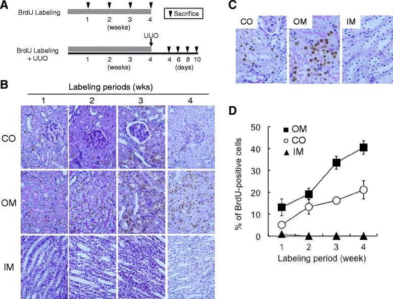
Fig. 1.

Presence of BrdU-positive cells in the normal rat kidneys after long-term BrdU labeling. a Experimental design. BrdU was intraperitoneally injected into normal rats for the indicated periods. After BrdU labeling, the rats were sacrificed, and the kidneys were removed and used for histological examination. In another experiment, UUO was induced in rats pre-treated with BrdU for 4 weeks as described in “Methods”. At the indicated times after UUO, the rats were sacrificed and the kidneys were removed and used for histological examination. b, c Detection of BrdU-positive cells in the normal rat kidneys after long-term BrdU labeling. BrdU was injected intraperitoneally into the normal rats for the indicated periods. BrdU-positive cells (brown nuclei) were examined by immunostaining and were counterstained with PAS. Magnification is ×400 in b and ×1000 in c. CO cortex, OM outer medulla, IM inner medulla. d Quantitative analysis of BrdU-positive cells. (Filled square) OM, outer medulla, (empty circle) CO, cortex, (filled triangle) IM, inner medulla. Values are means ± SE (n = 5)
