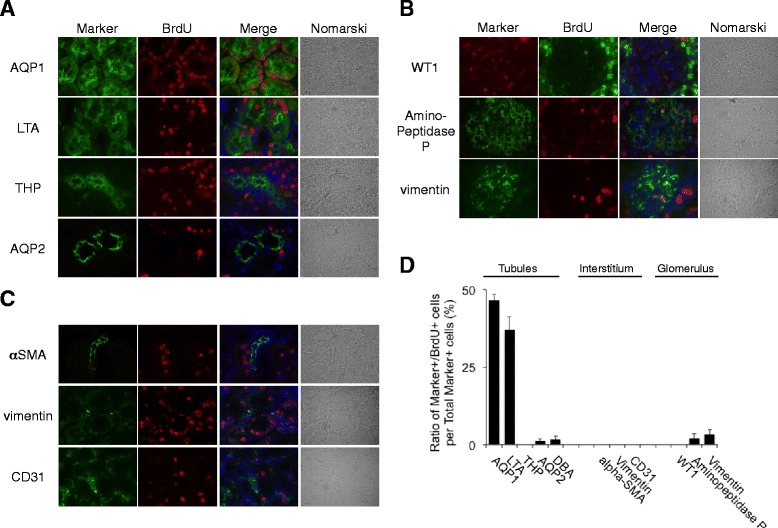
Fig. 2.

Localization of BrdU-positive cells in the normal rat kidneys after long-term BrdU labeling. a–c BrdU was given intraperitoneally to normal rats for 4 weeks. Double staining of BrdU with several tubular (a), glomerular (b), and interstitial (c) markers was performed. Markers: aquaporin-1 (AQP-1), lotus tetragonolobus agglutinin (LTA), Tamm-Horsfall glycoprotein (THP), aquaporin-2 (AQP-2), alpha-SMA, vimentin, CD31, WT1, and aminopeptidase P. DAPI (blue). Magnification, ×1000. d Quantitative analysis of percentage of BrdU-positive cells per total marker-positive cells. Values are means ± SE (n = 5)
