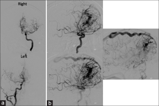
Figure 2.

(a) Preembolization digital subtraction angiograms of the anterior circulation performed by injection of the left internal carotid artery and right internal carotid artery demonstrating vascular pedicles emerging from the right middle cerebral artery and right anterior communicating artery (ACA) (top), and pedicles emerging from the left ACA (bottom). (b) Mid to late phase angiogram demonstrate the arteriovenous malformation nidus and the venous outflow
