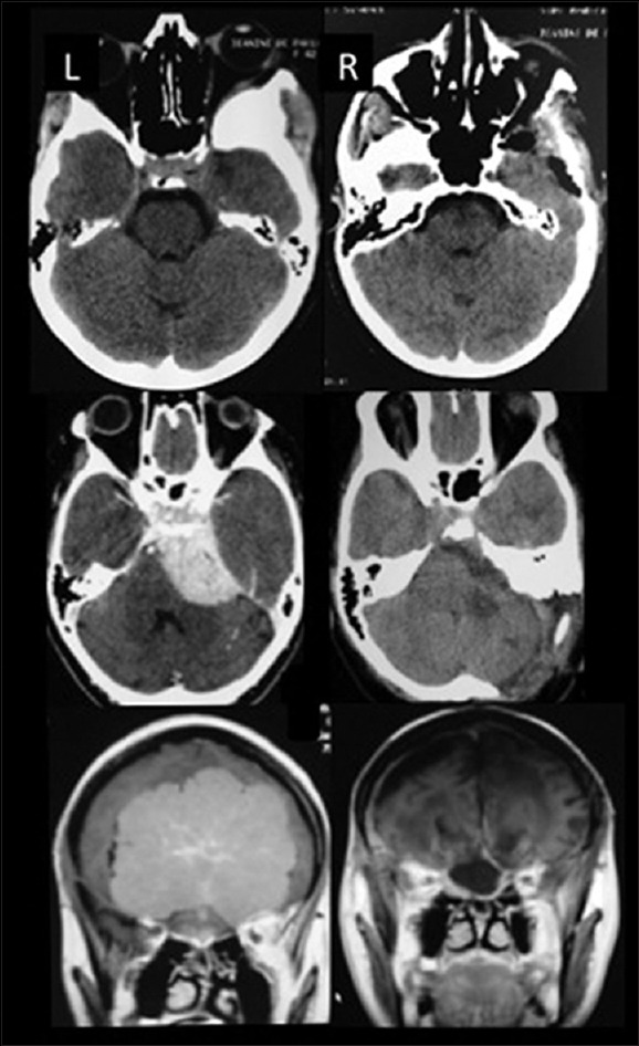
Figure 2.

Illustrative cases – Upper: Computed tomography (CT) scan sphenoorbital meningioma, Middle: CT scan petroclival meningioma, Lower: Magnetic resonance imaging olfactory groove meningioma. L (left): Preoperative images, R (right): Postoperative images. Observe the aggressive bone removal in all three cases
