Fig. 6.

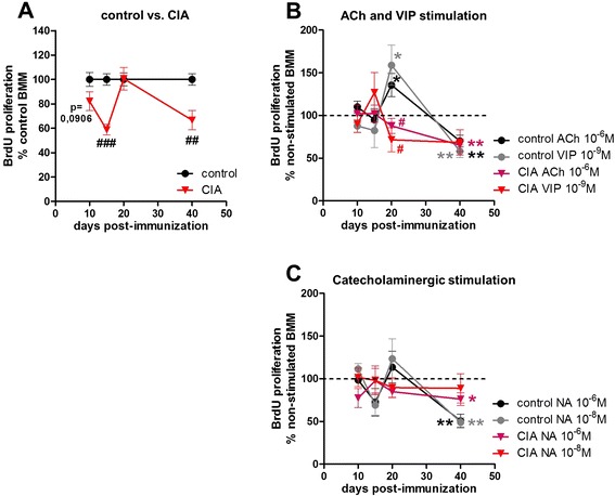
BrdU proliferation assay of pre-cultured BMMs. Pre-cultured BMMs from control and CIA animals which were serum-deprived for 24 h were cultured in the presence of BrdU for 48 h. Percentage of cell proliferation of BMMs from CIA compared with control animals is depicted in (a). Influence of ACh and VIP on proliferation of BMMs from control and CIA rats is shown as percentage to non-stimulated macrophages (non-stimulated = dotted line) (b). Respective results for noradrenergic stimulation are shown under (c). N (control/CIA) = day 10 (11/12), day 15 (12/12), day 20 (12/12), and day 40 (14/14). Neurotransmitter stimulation N (control/CIA) = day 10 (6/6), day 15 (6/6), day 20 (8/8), and day 40 (8/8) for ACh, VIP, and NA (10−6 M, 10−8 M). Data are expressed as mean ± standard error of the mean. *P < 0.05; **P < 0.01 neurotransmitter stimulation versus non-stimulated cells # P < 0.05; ## P < 0.01; ### P < 0.001 controls versus CIA. ACh acetylcholine, BMM bone marrow-derived macrophage, BrdU 5-bromo-2′-deoxyuridine, CIA collagen II-induced arthritis, NA noradrenaline, VIP vasoactive intestinal peptide
