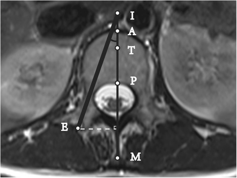
Fig. 2.

Cross-sectional MRI scan of lumbar pedicle: Unsuccessful puncture at the Max-angle. Line M: Anteroposterior midline of the vertebra; A: the intersection point of Line M and the vertebral anterior edge; P: the intersection point of Line M and the vertebral posterior edge; T: the anterior trisection point of Line AP; ∠EIP: Puncture Max-angle; Line EI was used to simulate the puncture device, which was 3.5 mm in diameter; the inner edge of Line EI was tangential to the medial wall of the pedicle, and the outer edge of Line EI was tangential to the lateral wall. In Fig. 2, point I was on the extension line of AP, and AI was defined as a negative value. The Puncture Success Value = 100*AI/AP < 34, and the puncture was considered a failure
