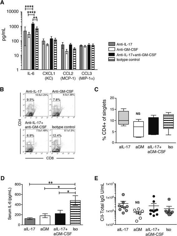
Fig. 2.

Cytokines in synovial washouts and serum and flow cytometric analysis of synovium and serum immunoglobulin G (IgG) after 14 days of interleukin (IL)-17 and granulocyte macrophage colony-stimulating factor (GM-CSF) blockade. a Luminex analysis of cytokines and chemokines in joint washouts (n = 6 mice per group). b Flow cytometric analysis of phorbol 12-myristate 13-acetate/ionomycin-stimulated synovial tissue stained with monoclonal antibodies for CD4 and CD8 after 14 days of treatment. The gate depicts the percentage of CD4+ cells of single synoviocytes. Plots shown are representative of n = 6 joints/group. Mean ± standard error of the mean (SEM) for each group is given in the fluorescence-activated cell sorting plot. c Summary graph for the flow cytometric analysis of synovial tissue. Box depicts 25th to 75th percentiles. Line depicts median. Whiskers depict minimal to maximal values. n = 6 mice per group. d IL-6 levels in serum measured by Luminex assay. n = 10 mice/group. Mean ± SEM. e Total collagen-specific immunoglobulin G (IgG) in serum. n = 7–10 mice/group. *p < 0.05; **p < 0.01; analysis of variance followed by Bonferroni’s test for multiple comparisons. CCL CC chemokine ligand, CII type II collagen, CXCL chemokine (C-X-C motif) ligand, KC keratinocyte, MCP monocyte chemoattractant protein, MIP macrophage inflammatory protein, NS not significant
