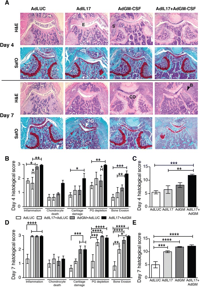
Fig. 3.

Histological analysis of joint damage after intraarticular injection of adenoviral vectors for interleukin (IL)-17 and granulocyte macrophage colony-stimulating factor (GM-CSF). a Haematoxylin and eosin (H&E)- and Safranin O (SafO)-stained sections of knee joints on days 4 and 7 after adenoviral transfer. E exudate, S synovitis, CD cartilage damage, B bone erosion, PG proteoglycan depletion. Representative sections are shown from n = 6 joints/group. b Individual and c total histological scores for day 4 after adenoviral transfer. d Individual and e total histological scores for day 7 after adenoviral transfer. Mean ± standard error of the mean for n = 6 joints per group. *p < 0.05; **p < 0.01; ***p < 0.001; ****p < 0.0001; analysis of variance followed by Bonferroni’s test for multiple comparisons
