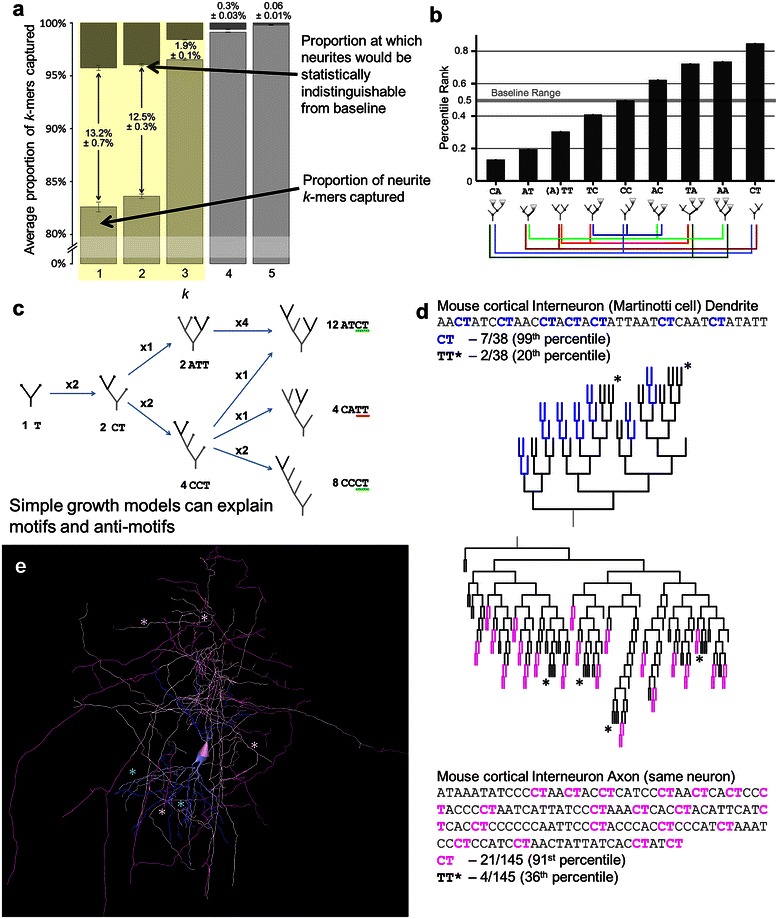
Figure 5.

Dimer analysis reflects terminal growth effects. a. Average proportions of captured k-mers (percentile rank between 2.5th and 97.5th) at each k for all neurites, with a break between 0 and 80%. Over 95% of trimers are captured, as are over 98% of tetramers, suggesting that most analyses should focus up to dimers, and possibly trimers. Darker descending bars represent the baseline, or chance level based on bootstrapping of surrogates. Numbers between or above bars signify the gap in percent captured between real neurites and the baseline (i.e. statistical equivalence). b. All axon and dendrite dimers except CC are motifs or anti-motifs. Colored lines below dimer schematics show associations between dimers. When one dimer proportion increases (and in turn its percentile rank) another must decrease. c. Growth processes produce specific topological patterns. Starting with a tree of a single bifurcation, the tree grows one bifurcation at a time from terminal nodes. After two bifurcation steps the CT to TT ratio is 2:1 as there are twice as many ways a CCT shape can emerge compared to an ATT shape. After a third bifurcation (at right), the dimer ratio is 5:1, and still 3:1 controlling for %C. d and e. CT and TT motifs and anti-motifs shown in an exemplar interneuron’s dendritic and axonal arbors (NMO_00340 from [68]) in sequence and dendrogram (d), and full morphology (e). Sequence and dendrogram highlighting indicate CT dimers in the axon (pink) and dendrite (blue). In the morphology, the darker color indicates the CT dimers. Asterisks (*) indicate the TT dimer in both representations. All error bars are standard error of the mean.
