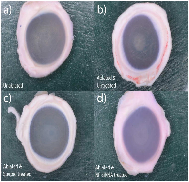
Figure 1. Visual comparison of ablated, unablated and treated corneas.
Freshly obtained rabbit globes were ablated to a depth of 155μm and cultured at the air-liquid interface for 14 days in serum free media supplemented with 1ng/ml of TGFB1. Four groups with six corneas each were tested experimentally – a) Unablated corneas, b) Laser ablated with no treatment, c) Laser ablated and treated with Steroids, d) Laser ablated and treated with triple siRNA complexed with nanoparticles.

