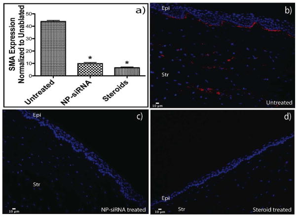
Figure 6. RNA analysis and Immunohistostaining of corneal tissues show reduction in SMA after treatment with steroids and NP-siRNA.
RNA from the scar-like tissue was collected using a 8mm punch biopsy and the expression levels of SMA were analyzed with qRT PCR. 18S rRNA was used as the housekeeping gene and all expression levels were normalized to their respective levels in unablated corneas. Image a shows the reduction in SMA expression in treated corneas when compared to the ablated corneas. For immunohistostaining, corneas from the experimental groups were fixed overnight in 4 % paraformaldehyde. They were then bisected, embedded in OCT and then sectioned in 10μm slides. To stain for SMA, slides were blocked in horse serum and then incubated with cy3 labeled SMA antibody. Rabbit globes that were ablated and untreated show SMA staining in the basal epithelium and stroma (Figure a). Figure b) and c) show higher magnification of areas with myofibroblasts. Globes that were ablated and treated with NP-siRNA and steroids show no SMA staining (Figures d and e).

