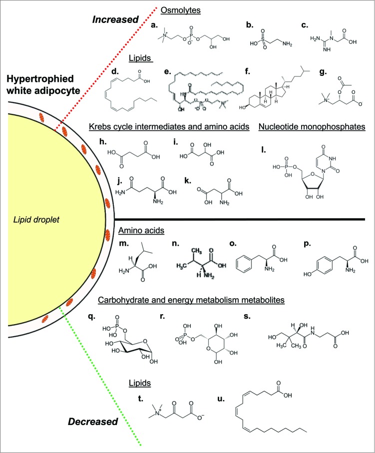
Figure 2.
Overview of biochemical changes occurring during adipocyte whitening. Conditions of chronic nutrient excess leading to adipocyte hypertrophy are associated with changes in several metabolic pathways. Increased in abundance (top; a-l) are several osmolytes including glycerophosphorylcholine (a), taurine (b) and hypotaurine (not shown), and creatine (c). Also increased are numerous fatty acids. Shown is arachidonate (d) as well as its downstream product 12-HETE (not shown), which may be involved in adipocyte inflammation. Other indicators of adipocyte inflammation that were higher in abundance in adipose tissues from obese mice include stearoyl sphingomyelin (e) and cholesterol (f). Evidence of lipid buffering is suggested by elevated levels of the carnitine lipid derivatives acetylcarnitine (g) and propionylcarnitine (not shown). Several Krebs cycle intermediates and amino acids were increased in abundance as well; these include succinate (h), malate (i), glutamine (j), glutamate (not shown), and aspartate (k). Additionally, the nucleotide monosphosphates uridine monophosphate (l), cytidine monophosphate (not shown) and inosine monophosphate (not shown) were increased. Also increased in obese mice were the 6-carbon sugars glucose and maltose (not shown). In tandem with the higher levels of these metabolites were decreases in numerous other biochemicals. Decreases in several amino acids involved in anaplerosis, including leucine (m), valine (n), phenylalanine (o) and tyrosine (p), were observed in adipose tissues of high fat-fed mice. Levels of phosphorylated glucose (q) and mannose (r) as well as pantothenate (s), which is important in coenzyme A synthesis, were diminished as well. Finally, the carnitine degradation product 3-dehydrocarnitine (t) and mead acid (u) were metabolites in the lipid family that were lower in abundance in adipose tissues from high fat-fed mice.

