Fig. 7.

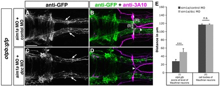
Dcc-mediated attraction causes medial displacement of otpb:gfp longitudinal axons after sim1a knockdown. Dorsal views of hindbrain confocal z-projections of 48 hpf otpb:gfp embryos. Anterior is towards the left. (A-D) GFP-positive longitudinal axons are medially displaced towards the midline (A, arrows) after co-injection of sim1a MO and control MO in otpb:gfp embryos. Medial displacement of longitudinal otpb:gfp axons (C, arrows) is reduced after combined injection of sim1a MO and dcc MO. (E) Quantification of the distance between otpb:gfp-positive longitudinal axons (I) or of the distance between Mauthner neurons (II) of indicated treatments. Arrowheads in B,D indicate normal midline crossing of MA neurons. *P<0.001; n.s., not significant. Scale bar: 50 μm.
