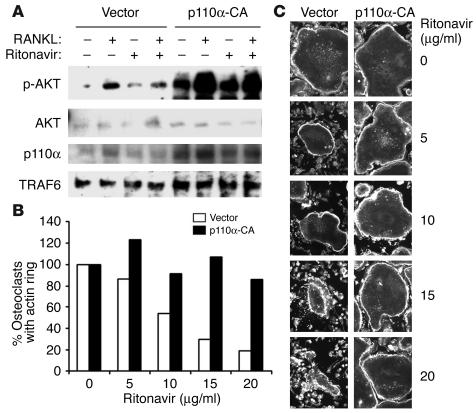
Figure 7.
Introduction of PI3K-CA restores RANKL-induced phosphorylation of Akt and osteoclast actin ring formation in the presence of ritonavir. (A) Retroviral transduction of either vector or PI3K-CA (p110a-CA) into bone marrow macrophages was followed by 3 days of culture in selection media, M-CSF, and RANKL. After starvation (3 hours) and pretreatment (1 hour) with either vehicle or ritonavir, cells were stimulated with RANKL for 15 minutes. Immunoblots reveal restoration of RANKL-induced Akt phosphorylation when PI3K-CA is introduced. As expected, total PI3K is enhanced as a result of transduction (p110α blot). TRAF6 Western blots act as a loading control. (B) Percentage of osteoclasts with intact actin rings after ritonavir exposure is quantitated. (C) Osteoclasts, retrovirally transduced with either vector or PI3K-CA, were generated on glass coverslips. After 4 days, cells were exposed to various doses of ritonavir for 2 hours, then processed for immunofluorescence microscopy for β-actin. Dose-dependent disruption of the characteristic actin ring of the osteoclast cytoskeleton is observed in vector but not PI3K-CA–transduced cells.

