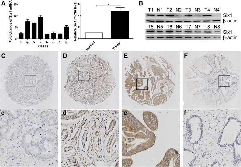
Fig. 1.

Expression of Six1 in prostate cancer tissues and normal prostate tissues detected by western blotting and immunochemistry. a Relative mRNA expression of Six1 normalized to β-actin was calculated (n = 8), ★ indicates P < 0.05. b Expressions of Six1 protein in 8 paired tissues were examined by western blotting. Micrographs showed weak (c, c), moderate (d, d), and strong (e, e) staining of Six1 in prostate cancer tissues, as well as low (f, f) expression of Six1 in normal prostate tissues (upper magnification × 100, lower panel: magnification × 400.)
