
Figure 4. Visualization of loss of kinase activity of cdk2/cyclin E in the presence of peptides TAALS and LAALS via (γ-32P) ATP labeling on the CTD of RNAPII.
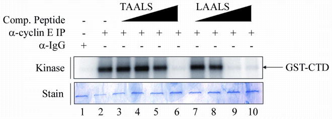
Immunoprecipitation of cyclin E in the presence of peptides was performed as in the Western blot analysis. Peptides TAALS and LAALS were added in increasing concentrations (0.01, 0.1, 1, and 10μM) to the kinase reaction. Kinase activity of cdk2/cyclin E was monitored via the addition of GST-CTD as a substrate. Dissociation of the cdk2/cyclin E complex is monitored by the decrease in phosphorylation of the CTD of RNAPII. Peptide LAALS required a lower concentration in order to exhibit a certain level of inhibition of kinase activity in vitro as compared to peptide TAALS. The lower panel shows the stained gel indicating equal loading of substrate among all lanes.